Architects in Lucknow for Luxury Homes, Interiors & Construction
Design to handover, one team
Hasan & Kushal Architects designs private villas, bungalows and premium workspaces across Lucknow. We take your project from the first sketch through approvals, interiors and construction, so one team is answerable for the result.
About the firm
An Architecture Firm in Lucknow Built Around Premium Homes
Project Completed
0+
Years of Expereience
0+
Sq. Ft. Designed
0K+
Cities Served
0+
Hasan & Kushal Architects is an architecture firm in Lucknow founded and led by two partner architects, Ar. Hasan Zaidi and Ar. Kushal Dubey, working from Gomti Nagar. Both partners stay involved in every project, so you get two experienced architects reviewing your plan, not one. We design homes and workspaces for clients who want the plan, the interiors and the build to feel like one idea, not three separate contracts.
Every project starts with how you live or work: your plot, your family, your daily routine, your budget.
What we do
Architect and Interior Designer in Lucknow, Under One Roof
Most homeowners in Lucknow hire an architect, then an interior designer, then a contractor, and spend months coordinating them. We bring all three together.
Architectural Design
Luxury villas, independent bungalows, offices and commercial buildings. Site study, planning, elevations, 3D views and working drawings.
Interior Design
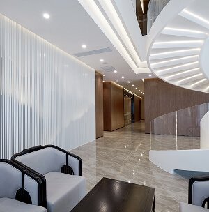
Premium interiors for homes and offices: space planning, false ceilings, lighting, modular kitchens, furniture and material selection, all drawn to match the architecture.
Turnkey Construction
From foundation to finishing, built by our team under the architect’s supervision, with a fixed scope and a clear schedule.
Not sure where to start? Bring your plot papers. We’ll tell you what’s possible.
A contemporary luxury home in Aliganj with a premium front elevation and clean architectural lines. A double-height entrance porch, spacious balconies and terraces, and a landscaped front lawn bring the design together.
Architecture and interiors for a family home in Mahanagar, from floor layouts and a modern elevation to living, dining, bedroom and kitchen design. Planning for natural light and ventilation runs through every room.
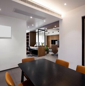
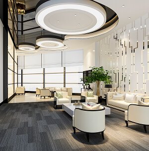
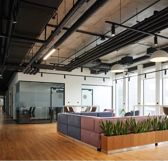
A complete office interior with executive cabins, meeting rooms and collaborative workstations. Custom furniture, a premium reception, false ceilings with ambient lighting and glass partitions keep the space professional and comfortable.
We visit your plot, study its orientation and bylaws, and note how your family wants to live.
Concept and budget
02
You get layout options, 3D views and a budget range before any detailed drawing starts.
Design and approvals
03
Working drawings, structure, services and interiors are finalised.
Build and handover
04
Construction moves ahead on site with regular progress updates until the keys are in your hand.
Client stories
What Our Clients in Lucknow Say
4.8/5
Whether you’re just beginning your fitness journey or pushing toward new personal records, our state-of-motivating is a atmosphere, we’re here to help you unlock your full potential. From weight training and the cardio to group classes and functional fitness zones, every detail is designed to elevate your performance and well-being. Join us and discover what your body and mind are truly capable of.
Saad Alam
Company Owner
4.8/5
Whether you’re just beginning your fitness journey or pushing toward new personal records, our state-of-motivating is a atmosphere, we’re here to help you unlock your full potential. From weight training and the cardio to group classes and functional fitness zones, every detail is designed to elevate your performance and well-being. Join us and discover what your body and mind are truly capable of.
Abu Talha
Manager
FAQS
Questions People Ask Before Hiring an Architect in Lucknow
Still have a question?
Talk to an architect, not a call centre
Tell us about your plot or project and we will answer your questions directly, with no obligation.
Fees depend on the built-up area, the type of project and the scope you need, whether that is design only or design, interiors and construction together. After a site visit we give you a clear written quote, so there are no surprises later.
Yes. You can hire us for architecture only, interiors only, or the full package. Most clients choose the full package because one team stays answerable for the result.
Yes. Along with architecture and interiors, we manage premium construction, so design intent, quality and timelines stay with one accountable team from the first drawing to handover.
Concept layouts and 3D views usually take a few weeks, followed by working drawings, structure and services. We agree a clear timeline with you at the start, based on the size and scope of your project.
Yes. We prepare the drawings needed for map approval and design your home within the applicable bylaws, so the approval process goes as smoothly as possible.
Your plot papers or site plan, a rough budget, and photos of homes you like. If you don’t have these yet, we can still meet and talk through what’s possible.
AREAS WE SERVE
Your Architect in Lucknow, Wherever You Are Building
Hasan & Kushal Architects is an architect in Lucknow working across the city, from compact independent houses to large villas, offices and commercial buildings. Because we are based here, we know local plot sizes, LDA and Awas Vikas bylaws, soil and climate, and the contractors and suppliers who deliver quality on site.
Wherever your plot is in Lucknow, our team visits in person, studies the site and gives you a clear plan and budget before design begins.